|
Healthy Living...
|
||||||||||||


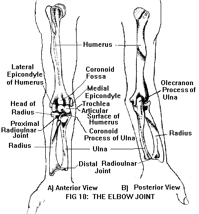
ELBOW (see also Anatomy of the Joints)The elbow joint (Figure 10) is formed by the articulation of the humerus (upper arm bone) with the radius and ulna (the two bones of the forearm). (The radius is located on the thumb side of the forearm, the ulna on the little finger side.) The elbow is a hinge-like joint, capable of two movements: flexion (bending) and extension (straightening). Just below the elbow hinge, the radius and ulna articulate with each other to form the proximal radioulnar joint. (There is also a distal radioulnar joint, just above the wrist.) The radioulnar joints permit rotation of the forearm so that the palm faces upward (supination) or downward (pronation).
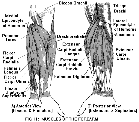
Movements of the elbow (flexion/extension) and forearm (pronation/supination) are carried out by different muscle groups. Flexion of the elbow is produced by three muscles: biceps brachii, brachialis, and brachioradialis. In addition to elbow flexion, the biceps brachii supinates the forearm (with the help of another muscle, called supinator). Extension of the elbow is produced by two muscles: triceps brachii and anconeus. Pronation of the forearm is carried out by two muscles: pronator quadratus and pronator teres. The lower end of the humerus flares out into two rounded projections (one on each side of the elbow) called epicondyles. The lateral epicondyle is on the outer side; the medial epicondyle is on the inner side. These serve as muscle attachments. The medial epicondyle is the attachment for the pronator teres muscle, as well as for a group of wrist and finger flexors. The lateral epicondyle is the attachment for supinator, as well as for a group of wrist and finger extensors. The muscles of the forearm, and their epicondylar attachments are shown in Figure 11.
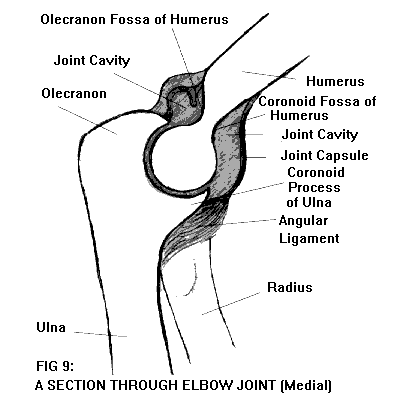
The upper end of the ulna is shaped somewhat like a crescent, with two projections (Figure 9). The uppermost projection is called the olecranon; it forms the point of the elbow. When the elbow is extended, the olecranon fits into a depression (the olecranon fossa) on the posterior surface of the humerus. The other projection of the crescent is called the coronoid process; when the elbow is flexed, it fits into another depression (the coronoid fossa) on the anterior surface of the humerus.
The disc-shaped upper end of the radius is called the head. The radial head articulates with the humerus, and also with the coronoid process of the ulna. The elbow joint is bound together by several ligaments. The ulnar collateral ligament stabilizes the medial side of the joint; the radial collateral ligament stabilizes the lateral side of the joint. The annular ligament encircles the head of the radius, binding it to the ulna. The three major nerves of the upper extremity (the radial, ulnar, and median nerves) pass through the elbow region on their way to the hand. The ulnar nerve passes behind the medial epicondyle, close to the surface. Bumping or leaning on the elbow may compress the ulnar nerve (the so-called "funny bone"), producing tingling or numbness on the ulnar (little finger) side of the hand.
|

_______________
Stop Letting Elbow Pain Limit YOU!ģ.. Your
Risk Free Opportunity to Become Pain Freeģģ ______________
No More TMJD _____________
_______________ Scientifically Proven Shin Splints Treatment |